
Forgetting is as much a part of recalling memories, stories and emotions as remembering itself. But while we sometimes forget our keys, a name or even an important date, this forgetfulness can occasionally indicate something far more serious and urgent. When it comes to diseases as debilitating as Alzheimer’s and other forms of dementia, scientists have long been curious to understand what actually happens in the brain.Now, researchers at Johns Hopkins University, in a study published in the journal NeuroImage: Reports in September 2026, have identified changes in two brain regions that could offer an important clue, with their findings suggesting that these areas may begin to shrink years before a person experiences noticeable memory loss.According to a ScienceDaily report dated November 14, 2026, the researchers found that while the hippocampus is an important part of the brain examined for signs of Alzheimer’s, two other regions associated with memory and emotional processing could help scientists understand the development of the disease before the actual loss of memory becomes apparent.The changes could become detectable as much as five to ten years before the clinical symptoms of the disease emerge, potentially opening a new pathway for understanding its silent progression and exploring treatments during this period.
Two significant regions of the brain apart from the hippocampus
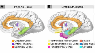
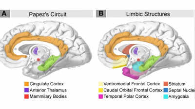
But before understanding this discovery, it is important to know what these different regions of the brain are responsible for. The hippocampus is particularly important when it comes to forming and consolidating memories, which is why damage to this area is often associated with the memory difficulties experienced by people with Alzheimer’s disease. But it is not the only part of the brain involved in these processes.Located near the hippocampus, the entorhinal cortex acts as an important part of the brain’s memory network, helping process and retrieve previously stored information. The amygdala, meanwhile, plays a major role in processing emotions and remembering experiences that carry an emotional association. Together, these regions contribute to the way people process, retain and recall information from their lives.This research also suggests that changes in these two areas may become apparent before the hippocampus shows comparable signs of deterioration. By examining the entorhinal cortex and amygdala alongside it, scientists may be able to identify signs of the disease that could otherwise go unnoticed during its earliest stages.
What the brain scans revealed
For the purpose of the scientific study, the researchers examined MRI and PET brain scans from two independent research cohorts: the Alzheimer’s Disease Neuroimaging Initiative (ADNI) and the Biomarkers of Cognitive Decline Among Normal Individuals (BIOCARD) study. These projects included people at different stages of Alzheimer’s disease, including those who had no noticeable cognitive symptoms and individuals experiencing cognitive impairment.By examining changes in the brain over time, the team found a consistent pattern across both groups. The entorhinal cortex and amygdala experienced greater relative tissue loss during the earlier stages of Alzheimer’s than the hippocampus.Abnormal tau protein buildup has also been found to play a significant role in the development of Alzheimer’s disease. Under normal circumstances, tau helps ensure that the internal structure of nerve cells remains stable. However, in people with Alzheimer’s, this protein can accumulate abnormally and form tangled structures inside brain cells, interfering with their functioning.Using PET imaging and digital pathology, the team found that areas showing signs of tissue loss also corresponded with regions displaying tau-related changes.
Could this help identify Alzheimer’s earlier?
One of the more promising aspects of the research is what it could mean for identifying the disease before its effects on everyday life become apparent. Alzheimer’s can develop over a long period, with biological changes taking place in the brain well before a person begins experiencing significant difficulties with memory, thinking or other cognitive abilities. By the time these symptoms become noticeable, considerable changes may already have occurred.Michael Miller, a biomedical engineer at Johns Hopkins University and the study’s senior author, explained that these changes could provide a window of five to ten years during which the brain is already showing signs of deterioration, even though clinical symptoms have not yet appeared. The possibility of recognising these changes earlier could eventually allow doctors to explore interventions before more extensive damage takes place.While the findings are a significant development in medical research, they should not be mistaken for a diagnostic breakthrough that is already ready for use in hospitals. Although the researchers have identified promising imaging markers, the team hopes to investigate the molecular changes taking place in these brain regions, especially those related to abnormal tau accumulation.With this study, a new pathway for further research into potential treatments has opened up, offering scientists more time to understand the processes responsible for significant tissue loss and potentially creating new avenues for treatment, especially for individuals with a family history of the disease.