
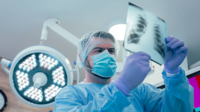
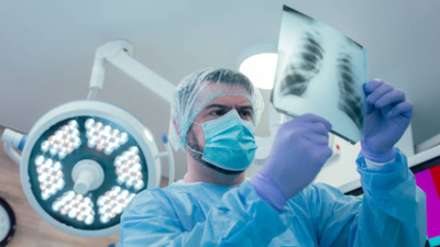
A persistent cough, chest discomfort and an unusual finding inside the chest cavity led doctors to an unexpected discovery: a living lung fluke. The parasite, which belongs to a group of worms capable of infecting human lungs, was found during a minimally invasive surgical procedure called thoracoscopy.The unusual case highlights a little-known parasitic infection called paragonimiasis, which can cause respiratory symptoms that resemble tuberculosis and other lung conditions.
What happened inside the patient’s chest?
The case was documented by researchers from China in a short communication published in October 2025 in Infection, Genetics and Evolution. The report, titled A case report of Paragonimus infection diagnosed by medical thoracoscopy, describes the woman who was admitted to hospital twice within six months because of recurrent pleural effusion, a condition in which fluid accumulates in the space between the lungs and chest wall.During her first hospital admission, blood tests using an enzyme-linked immunosorbent assay (ELISA) detected antibodies against two other parasites: Schistosoma japonicum and sparganum. However, the fluid accumulated again.When the woman returned to hospital, doctors decided to perform medical thoracoscopy to investigate the cause.That was when they spotted something moving. The parasite was contracting in a wave-like, peristaltic motion. It was identified under a microscope as an adult Paragonimus worm.Doctors also used metagenomic next-generation sequencing (mNGS) on the pleural fluid.The patient received another course of praziquantel. During four months of follow-up, doctors found no recurrence of the pleural effusion. The researchers described the case as the first documented visualisation of a living adult Paragonimus during thoracoscopy.
How does a lung fluke enter the human body?
Paragonimiasis is a food-borne zoonotic infection caused by parasitic flatworms belonging to the genus Paragonimus. Humans typically acquire the infection by eating raw or inadequately cooked freshwater crabs and crayfish carrying the infective larval stage of the parasite.Once swallowed, the larvae leave their protective cysts in the digestive tract, penetrate the intestinal wall and enter the abdominal cavity. From here they then migrate through body tissues, crossing the diaphragm and reaching the lungs.
Can the infection be treated?
Yes. Paragonimiasis is treatable with antiparasitic medicines.Diagnosis usually involves examining sputum or stool samples for parasite eggs. Blood antibody tests can also help, particularly during early infection, when the worms have not yet matured and started producing eggs. In unusual cases involving pleural disease, additional investigations, including thoracoscopy and molecular testing, may help establish the diagnosis.The researchers say their case demonstrates the diagnostic value of medical thoracoscopy and mNGS when conventional investigations do not establish the cause of recurrent pleural effusion.Prevention, however, begins much earlier, at the dining table.A parasite moving inside the chest may sound like an extraordinary medical discovery. But the more significant lesson from this case is that an infection acquired through contaminated food can remain difficult to identify even after repeated hospital visits.