
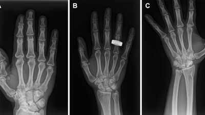
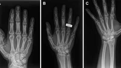
The first X-rays taken in space have shown that astronauts may soon have access to a medical tool that could become important on missions to the Moon and beyond. During the Fram2 mission in 2025, crew members used a digital X-ray system while orbiting Earth, producing images that researchers found diagnostically adequate. The achievement is significant because ultrasound has been the only reliable medical imaging method available during spaceflight for more than four decades. In the study published in Radiology, researchers tested whether a commercial, portable X-ray system could work in microgravity when operated by crew members with limited training. The results suggest that it can. Although the experiment was small and revealed practical difficulties with positioning, it showed that useful X-ray images could be captured without live ground support, opening a path towards better medical care far from Earth.
How did astronauts take the first X-ray images in orbit
The researchers tested a commercially available ‘portable digital radiography system’ during the 3.5-day Fram2 polar orbital mission. The mission launched on March 31, 2025, with four crew members aboard a Dragon spacecraft. The spacecraft travelled in a 90-degree polar orbit roughly 425 to 450 kilometres above sea level before returning to Earth on April 4. Three of the four crew members took part in the study. Importantly, none needed to become medical imaging specialists. According to the study, the participating crew members received about four hours of training before the flight.The system included an ultraportable wireless X-ray generator and a digital detector. Before launch, the equipment was tested for its ability to withstand the spacecraft environment, including vibration, temperature, vacuum and electromagnetic conditions. Once in orbit, the crew used the equipment without live ground support. They captured images of a hand, forearm, abdomen, pelvis and chest, as well as images of a phantom used to check image quality. They also X-rayed a smartwatch as part of testing whether the system could inspect equipment without taking it apart.
Why are X-rays important for astronauts during space travel
For astronauts travelling beyond Earth, a medical emergency could become difficult to assess quickly. On the International Space Station and other missions, ultrasound has traditionally been the main reliable imaging option. However, the researchers behind the study explained that ultrasound has limitations. It depends heavily on the person operating the equipment and requires suitable contact between the probe and the body. It can also be difficult to examine certain parts of the body. As missions become longer and move farther from Earth, having another way to look inside the body could become increasingly useful. X-rays offer a familiar and relatively fast way of examining bones and other structures. The challenge was finding out whether such imaging could actually work in the constantly moving environment of space.
Quality of the space X-ray images
The results were encouraging. The study found no evidence of meaningful differences between preflight and in-flight images in overall image quality, spatial resolution or contrast resolution. The average overall image-quality score for the seven in-flight anatomical radiographs was 4.86 out of 5, compared with 5 pre-flight ones. Spatial and contrast resolution also remained very similar. The biggest problem was not the X-ray equipment itself but positioning. Images of the chest, abdomen and pelvis were harder to position correctly in microgravity. The researchers reported significantly poorer positioning scores for central body images taken in flight. That difficulty makes sense in an environment where neither the astronaut nor the equipment naturally stays in one place. Even so, the researchers concluded that the images were diagnostically adequate.

Representative preflight, in-flight, and post-flight chest radiographs. Radiographs of the chest were acquired (A) preflight by a crew member, (B, C) in-flight on day 3 after launch (L+3) by a crew member, and (D) post-flight by a non-crew operator using the same imaging protocol. Image Credit: Radiological Society of North America(RSNA)
Could this help astronauts travelling to the Moon
The findings could become particularly relevant as human missions move farther from Earth. A short mission close to Earth can rely on rapid communication and support from medical teams on the ground. A mission to the Moon creates greater distance and operational challenges, while future journeys to Mars would make those problems even more serious. The study does not claim that X-rays are already ready for routine medical use on lunar or Martian missions. Instead, it demonstrates that the basic technology can work in orbit with relatively little crew training. The researchers also pointed out that the X-ray system could have uses beyond injuries and illness. During the Fram2 mission, X-rays were used to examine equipment internally. The technique allowed components to be viewed without opening or damaging them, with some internal features visible at a scale below one millimetre. That could eventually help crews investigate problems with spacecraft equipment, electronics or other mission hardware.
Challenges in front of the astronauts
The researchers identified several limitations. Only three crew members participated, and just two underwent X-ray imaging during the flight because of limited operational time. All were healthy and able-bodied, meaning the study could not fully reproduce the difficulties involved in imaging someone who was injured, in pain or unable to cooperate. Positioning also remains a major challenge. The study found that crew members needed better ways to align the X-ray source, detector and person being examined in microgravity.Another issue is communication. The images were reviewed on Earth as part of the research process, but the authors noted that real-time medical support may not always be possible during exploration missions. The equipment itself nevertheless performed well. After returning to Earth, the system was found to be functioning normally despite minor damage sustained during re-entry.
Future of space medical healthcare
The Radiology study describes the Fram2 experiment as the first acquisition of human radiographs in orbit. Its importance lies less in producing a single remarkable image and more in showing that a portable X-ray system can be operated by minimally trained crew members in microgravity. The researchers said that further improvements could make the equipment smaller, tougher and easier to use. With those changes, portable X-ray technology could eventually become part of medical equipment carried on future long-duration missions. For astronauts facing the risks of extended space travel, that could provide something Earth-based hospitals take for granted which is ‘another way to look inside the body when something goes wrong.’